English
 norsk
norskBlar i forfatter "Mao, Hong"
Viser treff 1-12 av 12
-
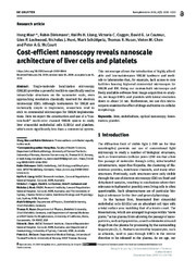
Cost-efficient nanoscopy reveals nanoscale architecture of liver cells and platelets
(Journal article; Tidsskriftartikkel; Peer reviewed, 2019-07-09)Single-molecule localization microscopy (SMLM) provides a powerful toolkit to specifically resolve intracellular structures on the nanometer scale, even approaching resolution classically reserved for electron microscopy (EM). Although instruments for SMLM are technically simple to implement, researchers tend to stick to commercial microscopes for SMLM implementations. Here we report the construction ... -
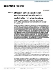
Effect of caffeine and other xanthines on liver sinusoidal endothelial cell ultrastructure
(Journal article; Tidsskriftartikkel; Peer reviewed, 2023-08-17)Xanthines such as caffeine and theobromine are among the most consumed psychoactive stimulants in the world, either as natural components of cofee, tea and chocolate, or as added ingredients. The present study assessed if xanthines affect liver sinusoidal endothelial cells (LSEC). Cultured primary rat LSEC were challenged with xanthines at concentrations typically obtained from normal consumption of ... -
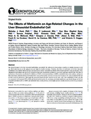
The Effects of Metformin on Age-Related Changes in the Liver Sinusoidal Endothelial Cell
(Journal article; Tidsskriftartikkel; Peer reviewed, 2020-06-14)Age-related changes in the liver sinusoidal endothelium, particularly the reduction in fenestrations, contribute to insulin resistance in old age. Metformin impacts on the aging process and improves insulin resistance. Therefore, the effects of metformin on the liver sinusoidal endothelium were studied. Metformin increased fenestrations in liver sinusoidal endothelial cells isolated from both young ... -
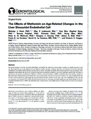
The effects of metformin on age-related changes in the liver sinusoidal endothelial cell
(Journal article; Tidsskriftartikkel; Peer reviewed, 2019-06-14)Age-related changes in the liver sinusoidal endothelium, particularly the reduction in fenestrations, contribute to insulin resistance in old age. Metformin impacts on the aging process and improves insulin resistance. Therefore, the effects of metformin on the liver sinusoidal endothelium were studied. Metformin increased fenestrations in liver sinusoidal endothelial cells isolated from both young ... -
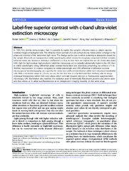
Label-free superior contrast with c-band ultra-violet extinction microscopy
(Journal article; Tidsskriftartikkel; Peer reviewed, 2023-03-03)In 1934, Frits Zernike demonstrated that it is possible to exploit the sample’s refractive index to obtain superior contrast images of biological cells. The refractive index contrast of a cell surrounded by media yields a change in the phase and intensity of the transmitted light wave. This change can be due to either scattering or absorption caused by the sample. Most cells are transparent at visible ... -
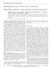
Manipulating fenestrations in young and old liver sinusoidal endothelial cells
(Journal article; Tidsskriftartikkel; Peer reviewed, 2018-12-27)Fenestrations are pores within liver sinusoidal endothelial cells (LSECs) that enable the transfer of substrates (particularly insulin and lipoproteins) between blood and hepatocytes. With increasing age, there are marked reductions in fenestrations, referred to as pseudocapillarization. Currently, fenestrations are thought to be regulated by vascular endothelial growth factor and nitric oxide (NO) ... -
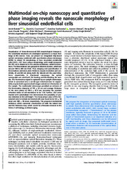
Multimodal on-chip nanoscopy and quantitative phase imaging reveals the nanoscale morphology of liver sinusoidal endothelial cells
(Journal article; Tidsskriftartikkel; Peer reviewed, 2021-11-23)Visualization of three-dimensional (3D) morphological changes in the subcellular structures of a biological specimen is a major challenge in life science. Here, we present an integrated chip-based optical nanoscopy combined with quantitative phase microscopy (QPM) to obtain 3D morphology of liver sinusoidal endothelial cells (LSEC). LSEC have unique morphology with small nanopores (50-300 nm in ... -
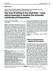
New ways of looking at very small holes – using optical nanoscopy to visualize liver sinusoidal endothelial cell fenestrations
(Journal article; Tidsskriftartikkel; Peer reviewed, 2018-01-10)Super-resolution fluorescence microscopy, also known as nanoscopy, has provided us with a glimpse of future impacts on cell biology. Far-field optical nanoscopy allows, for the first time, the study of sub-cellular nanoscale biological structures in living cells, which in the past was limited to electron microscopy (EM) (in fixed/dehydrated) cells or tissues. Nanoscopy has particular utility in the ... -
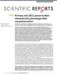
Primary rat LSECs preserve their characteristic phenotype after cryopreservation
(Journal article; Tidsskriftartikkel; Peer reviewed, 2018-10-02)Liver disease is a leading cause of morbidity and mortality worldwide. Recently, the liver non-parenchymal cells have gained increasing attention for their potential role in the development of liver disease. Liver sinusoidal endothelial cells (LSECs), a specialized type of endothelial cells that have unique morphology and function, play a fundamental role in maintaining liver homeostasis. Current ... -
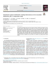
Quantitative analysis methods for studying fenestrations in liver sinusoidal endothelial cells. A comparative study
(Journal article; Tidsskriftartikkel; Peer reviewed, 2021-07-28)Liver Sinusoidal Endothelial Cells (LSEC) line the hepatic vasculature providing blood filtration via transmembrane nanopores called fenestrations. These structures are 50−300 nm in diameter, which is below the resolution limit of a conventional light microscopy. To date, there is no standardized method of fenestration image analysis. With this study, we provide and compare three different approaches: ... -
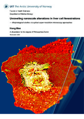
Unraveling nanoscale alterations in liver cell fenestrations - Morphological studies via optical super-resolution microscopy approaches
(Doctoral thesis; Doktorgradsavhandling, 2021-01-22)The endothelium makes up the innermost cell layer of blood vessels. It consists of a thin layer of simple squamous cells, forming an interface between circulating blood and the surrounding tissue. Endothelial cells of different vascular beds are specialized according to tissue-specific functions. For this project emphasis was placed upon high-resolution methods enabling the study of liver sinusoidal ...